We focus on applying biomechanical principles in clinical settings, particularly to improve the understanding, diagnosis, and treatment of neuromuscular disorders, orthopedic conditions, and age-related musculoskeletal issues. The primary motivation of our clinical biomechanics research is to enhance diagnostic tools and rehabilitation methods for musculoskeletal problems, where biomechanical approaches play a crucial role.
Neuromuscular Disorders
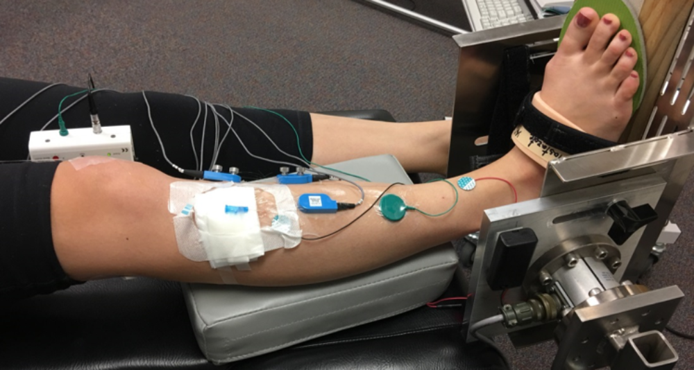
Neuromuscular diseases change musculoskeletal system as well as material properties of skeletal muscles. Our recent studies on patients with spastic cerebral palsy showed that understanding the sources of joint restrictions are not straight forward. Studies reporting adaptation for some of the muscles are not capable of explaining the limited joint range. Relating individual muscles’ mechanical characteristics to the joint motion restriction is crucial but more complicated. Driven by this gap in understanding, our goals are to reveal the origins of the motion and mobility limitations caused by musculoskeletal conditions such as cerebral palsy, stroke, muscular dystrophy, multiple sclerosis.
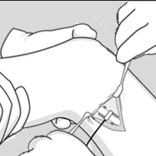
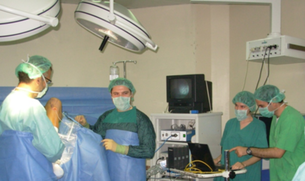
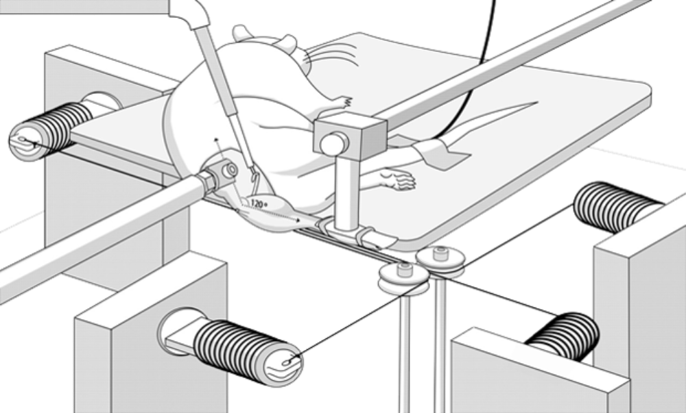
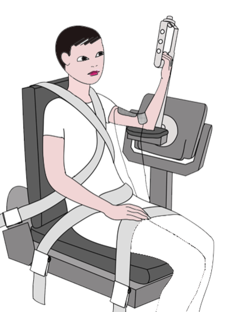
To do so, we aim to develop an extensive approach that combines invasive (e.g. intra-operative measurements), minimally invasive, and non-invasive (motion and mobility analysis, ultrasound based measurements, EMG, torque measurements, MRI) experimental methods with modeling and simulations. This approach would reveal the probable causes of knee, ankle, wrist, and elbow motion limitations as well as the effects of orthopedic interventions. Hence, the outcomes of our research would improve the treatment methods.
Publications:
Brendecke E., Tsitlakidis S, Götze M., Hagmann S. and Ates F., 2023: Quantifying the effects of achilles tendon lengthening surgery: An intraoperative approach. Frontiers in Physiology, 14:1143292.
Ateş F., Brandenburg J., Kaufman K.R., 2020: Effects of Selective Dorsal Rhizotomy on Ankle Joint Function in Patients with Cerebral Palsy. Frontiers in Pediatrics, 8.
Kaya C.S., Bilgili F., Akalan N.E., Temelli Y., Ateş F., Yucesoy C.A. 2019: Intraoperative experiments combined with gait analyses indicate that active state rather than passive dominates the spastic gracilis muscle’s joint movement limiting effect in cerebral palsy. Clin Biomech. 68: 151-157.
Ateş F., Temelli Y., Yucesoy C.A., 2018: Effects of Antagonistic and Synergistic Muscles’ Co-activation on Mechanics of Activated Spastic Semitendinosus in Children with Cerebral Palsy. Human Movement Science. 57: 103-110.
Kaya C.S., Ateş F., Temelli Y., Yucesoy C.A., 2017: Effects of inter-synergistic mechanical interactions on the mechanical behaviour of activated spastic semitendinosus muscle of patients with cerebral palsy. J Mech Behav Biomed Mater. 77: 78-84.
Yucesoy C.A., Temelli Y., Ateş F., 2017: Intra-operatively Measured Spastic Semimembranosus Forces of Children with Cerebral Palsy. J. Electromyography & Kinesiology. 36: 49-55.
Ateş F., Temelli Y., Yucesoy C.A., 2016: The Mechanics of Activated Semitendinosus are not Representative of the Pathological Knee Joint Condition of Children with Cerebral Palsy. J Electromyography and Kinesiology. 28: 130-6.
Akdeniz Z., Bayramiçli M., Ateş F., Özkan N., Yucesoy CA., Ercan F., 2015: The role of botulinum toxin type a induced motor endplates following peripheral nerve repair. Muscle & Nerve, 52(3): 412-8.
Yucesoy, C.A., Turkoglu A.N., Umur S., Ateş F., 2015: Intact muscle compartment exposed to botulinum toxin type A shows comprised intermuscular mechanical interaction. Muscle & Nerve, 51(1): 106-16.
Ateş, F., Temelli Y., Yucesoy, C.A., 2014: Intraoperative experiments show relevance of inter-antagonistic mechanical interaction for spastic muscle’s contribution to joint movement disorder. Clinical Biomechanics, 29(8): 943-9.
Ateş, F., Temelli Y., Yucesoy, C.A., 2013: Human spastic Gracilis muscle isometric forces measured intraoperatively as a function of knee angle show no abnormal muscular mechanics. Clinical Biomechanics, 28: 48-54.
Yucesoy, C.A., Arıkan Ö.E., Ateş F., 2012: BTX-A Administration to the Target Muscle Affects Forces of All Muscles Within an Intact Compartment and Epimuscular Myofascial Force Transmission, J Biomech Eng, 134: 111002-1-9.
Yucesoy, C.A., Ateş F., Akgün, U., Karahan, M., 2010: Measurement of human Gracilis muscle isometric forces as a function of knee angle, intraoperatively. J Biomech, 43 (14): 2665-2671.
Collaborations:
- Sebastian Wolf, Universitätsklinikum Heidelberg, Klinik für Orthopädie und Unfallchirurgie, Heidelberg, Germany.
- Sébastien Hagmann, Universitätsklinikum Heidelberg, Kinderorthopädie, Neuroorthopädie und Fußchirurgie, Heidelberg, Germany.
- Marco Götze, Universitätsklinikum Heidelberg, Klinik für Orthopädie und Unfallchirurgie, Heidelberg, Germany.
- Urs Schneider, Fraunhofer-Institut für Produktionstechnik und Automatisierung (IPA), Stuttgart, Germany.
- Alper Yaman, Fraunhofer-IPA, Stuttgart, Germany.
- Okan Avcı, Fraunhofer-IPA, Stuttgart, Germany.
- Oliver Röhrle, University of Stuttgart, Stuttgart, Germany.
- Yener Temelli, Istanbul Medical School, Istanbul University, Istanbul, Turkey.
- Can A. Yucesoy, Institute of Biomedical Engineering, Bogazici University, Istanbul, Turkey.
- Kenton R. Kaufman, Motion Analysis Laboratory, Orthopedic Research, Mayo Clinic, Rochester, MN, USA.
- Richard L. Lieber, Shirley Ryan AbilityLab, Chicago, IL, USA.
Rehabilitation and Treatment of Neuromuscular Conditions
Botulinum toxin type A is one of the applications to treat spastic or hyperactive muscles. It causes muscle paralysis by inhibiting acetylcholine release into the presynaptic cleft in the neuromuscular junction and decreases muscle tone. A consequence of decreased hyperactivity is increased joint range of motion. Therefore, aim is to improve joint function and gait. Our studies on animal muscles indicated more complicated and some contradictory effects of botulinum toxin administration such as increased passive forces, enhanced connective tissue content, and decreased muscle length range of force exertion. Therefore, the efficacy of the treatment including the mechanical consequences of botulinum toxin in long-term requires more detailed research.
Publications:
Kaya Keles C.S. and Ates F., 2022: Botulinum toxin intervention in cerebral palsy-induced spasticity management: projected and contradictory effects on skeletal muscles. Toxins, 14: 772
Ateş F. and Yucesoy C.A., 2018: Botulinum Toxin Type-A Affects Mechanics of Non-injected Antagonistic Rat Muscles. J Mech Behav Biomed Mater. 84: 208-216.
Yucesoy C.A. and Ateş F., 2017: BTX-A Has Notable Effects Contradicting Some Treatment Aims in the Rat Triceps Surae Compartment, Which are not Confined to the Muscles Injected. Journal of Biomechanics. 66: 78-85.
Akdeniz Z., Bayramiçli M., Ateş F., Özkan N., Yucesoy CA., Ercan F., 2015: The role of botulinum toxin type a induced motor endplates following peripheral nerve repair. Muscle & Nerve, 52(3): 412-8.
Ateş, F., Ozdeslik R.N., Huijing P.A., Yucesoy, C.A., 2013: Muscle lengthening surgery causes differential acute mechanical effects in both targeted and non-targeted synergistic muscles. Journal of Electromyography and Kinesiology, 23 (5): 1199-205.
Ateş, F. and Yucesoy, C.A., 2014: Effects of BTX-A on non-injected bi-articular muscle include a narrower length range of force exertion and increased passive force. Muscle & Nerve, 49 (6): 866-878.
Collaborations:
- Zeynep Akdeniz, Marmara University Medical School, Istanbul, Turkey.
- Can A. Yucesoy, Institute of Biomedical Engineering, Bogazici University, Istanbul, Turkey.
Orthopedic Biomechanics
- Biomechanical Analyses of Surgical Methods
In collaboration with clinicians and surgeons, we design and perform biomechanical protocols to e.g. test the strength and quality of available or newly developed surgical knots, mechanically and physiologically evaluate the healing process of a nerve or a tendon repair etc.
- Muscle Length Optimization
A novel tool for determining optimal muscle length is needed in the clinic: During free gracilis muscle transfer surgery performed to replace the lost function of the biceps brachii, it is crucial to optimize the length range of the transferred gracilis muscle. Similarly, for reverse shoulder arthroplasty surgery to treat rotator cuff tears, the function of the deltoid muscles is essential for the success of the surgery: While the prosthesis provides a stable fulcrum and increases the range of motion it typically strains the deltoid muscles. Deltoid tension should be determined during the surgery to achieve maximal function. We aim to develop specific designs for each muscle that muscle tension can be predicted before surgery, can be used during surgery to determine exact length and location, and if necessary, can be used after surgery for non-invasive follow-ups. This approach has also great potential to be applied to tendon transfer surgery performed to correct joint deficiency in patients with cerebral palsy.
Publications:
Ateş, F., Ozdeslik R.N., Huijing P.A., Yucesoy, C.A., 2013: Muscle lengthening surgery causes differential acute mechanical effects in both targeted and non-targeted synergistic muscles. Journal of Electromyography and Kinesiology, 23 (5): 1199-205.
Gereli A., Akgün U., Uslu S., Ağır I., Ateş, F., Nalbantoğlu U., 2014: The effect of organic silicon injection on achilles tendon healing in rats. Acta Orthop Traumat Turc, 48 (3): 346-54.
Zeytin K., Ciloglu N.S., Ateş F., Vardar Aker F., Ercan F., 2014: The effects of resveratrol on tendon healing of diabetic rats. Acta Orthop Traumat Turc, 48 (3): 355-362.
Karahan M., Akgun U., Turkoglu A.N., Nuran R., Ateş F., Yucesoy C.A., 2012: Pretzel Knot compared with standard suture knots, Knee Surg Sports Traumatol Arthrosc, 20(11): 2302-6.
Collaborations:
- Mustafa Karahan, Acıbadem Hospital, Istanbul, Turkey.
- Umut Akgün, Acıbadem University Medical School, Istanbul, Turkey.
- Arel Gereli, Acıbadem University Medical School, Istanbul, Turkey.
- Can A. Yucesoy, Institute of Biomedical Engineering, Bogazici University, Istanbul, Turkey.
- Kenton R. Kaufman, Motion Analysis Laboratory, Orthopedic Research, Mayo Clinic, Rochester, MN, USA.
Aging
Age-related muscle weakness, sarcopenia, and neuromuscular diseases increase the incidence of accidents gradually. Therefore, knowing the age-related changes of musculoskeletal system is essential for early diagnosis, rehabilitation, and accident prevention. We have two key research lines in addressing some of these open research questions. One is extensive understanding of mechanics and physiology of aging muscles. The second is to develop methodologies to reverse some of the aging processes. While we perform various experimental techniques from histology, imaging to animal experiments for the first, the latter includes interventional studies on human.
Publications:
Ates F., Marquetand J. and Zimmer M., 2023: Detecting age‐related changes in skeletal muscle mechanics using ultrasound shear wave elastography. Scientific Reports, 13:20062
Muscle weakness
Muscle weakness is one of the major problems that occurs under diverse conditions. E.g., myopathies that cause muscle weakness are not easy to diagnose with standard lab tests. We aim to develop diagnostic tools to follow the course of diseases such as myopathies.
Publications:
Zimmer M., Kleiser B., Marquetand J., and Ates F., 2023: Characterization of muscle weakness due to myasthenia gravis using shear wave elastography. Diagnostics 13 (6), 1108
Collaborations:
- Yasuo Kawakami, Waseda University, Tokyo, Japan
- Kenton R. Kaufman, Motion Analysis Laboratory, Orthopedic Research, Mayo Clinic, Rochester, MN, USA.

Filiz Ates
Dr.Head of Experimental Biomechanics Group